Skip to content
AR/VR and 3D Printing Intersections
The worlds of 3D printing and mixed reality viewing have collided, and the files that can be used to present 3D anatomical visualizations in each have a high degree of overlap. While the two may seem to be in direct competition, they can actually work in tandem to improve overall quality of care.
Computer aided design files have their origin in “solid modeling” of objects, but both 3D printing and AR/VR use surface files as a means of presenting anatomical visualizations. The rationale for this is that it allows radiologists to provide care teams with dimensions, findings, and impressions of volumes like a fast-growing tumor as the surface that separates the tumor from surrounding tissue.
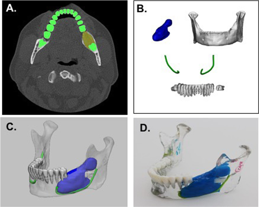
There is an entire science and art to presenting meaningful information in 3D, which is built into the software workflow suites radiologists use in hospitals every day. The figure below provides some basic visual insight into how a radiologist [A.] extracts the outlines of teeth, bones, tumors and nerves on CT slices through the body, [B.] defines the anatomy and pathology [the abnormal tumor in dark blue] regions of interests, [C.] presents in augmented reality, and how the same file mapped to [D.] appropriate printer materials can result in a tactile-visual investigation of a 3D print. For a more detailed description, please see this excellent peer-reviewed article by Dr. Nicole Wake and co-authors.

a Axial CT image showing segmentation of teeth (green) and tumor (yellow). b 3D anatomical regions of interest including the tumor (blue), mandible (white), teeth (white), and nerves (green). c 3D visualization of model including all anatomical parts. d 3D printed mandible tumor model including the mandible (clear), teeth (white), tumor (blue), and nerves (green)
Looking at the figure above, we can start to see how mixed reality and 3D printing can be synergistic or complementary, not in competition. Mixed reality is best for viewing. When visual communication is a priority, AR/VR provides almost instantaneous information transfer as opposed to waiting for a 3D print; indeed it may be sufficient on its own depending on user objectives. 3D printing is most amazing when tactile information and the ability to dissect and re-assemble parts using surgical practice tools, are the key objectives; for example, surgeries where the ability to feel and perceive is advantageous.
We can achieve synergy between the two methods quite easily. While waiting for the tactile print, you can view in AR/VR immediately and prepare your approaches while looking at the same model you will receive a physical version of hours, or even days, later.
There are many more ways that these technologies can work hand-in-hand when we consider use cases from communicating amongst clinical colleagues and teaching the next generation, to ‘getting your hands on the challenge ahead’ before surgery.
While the care team may desire a 3D print to communicate visually/tactilely, the value of mixed reality is being able to immediately convey ‘what is the problem, and what are we going to do about it’ both in today’s pandemic remote patient engagements [no visit required to see the model], and when we return to normal but want to preserve the efficiencies of some remote collaborations.
Interested in experiencing some of these efficiencies for yourself? Follow our blog links to repositories of files that you can try uploading to your iPhone today on our MedReality free trial. Then, once you find surface files like .STL, .WRL, .OBJ you want to download and try right now, consider how these technologies can not only co-exist, but also improve the care.
Get started bringing your medical models into AR here
Get unlimited uploads with MedReality Pro license here
Share This Story, Choose Your Platform!
