Skip to content
My Medical Adventure
Our VP of Clinical Mixed Reality, Colin Holmes, shares a personal experience of how visualizing a 3D model on MedReality helped educate and bring a level of comfort to his family during a scary incident…
For the past 20 odd years, I have been working with scientific visualization in one regard or another, initially with neuroimaging, then medical imaging and more recently, to using 3D visualization to improve the patient experience. Just prior to the Thanksgiving holiday, my family had a “medical adventure” which brought this work close to home. My daughter, a senior at a nearby private school, plays for their national caliber rugby team. While tackling a bigger girl she was struck on the back of the neck while being knocked over backwards to the ground where she hit the back of her head. Rugby is a full contact sport, and collisions like this are not infrequent, but in this case the trainer was concerned about the degree of neck pain and some other symptoms of head trauma, so we called for an ambulance to take her in for a detailed workup.
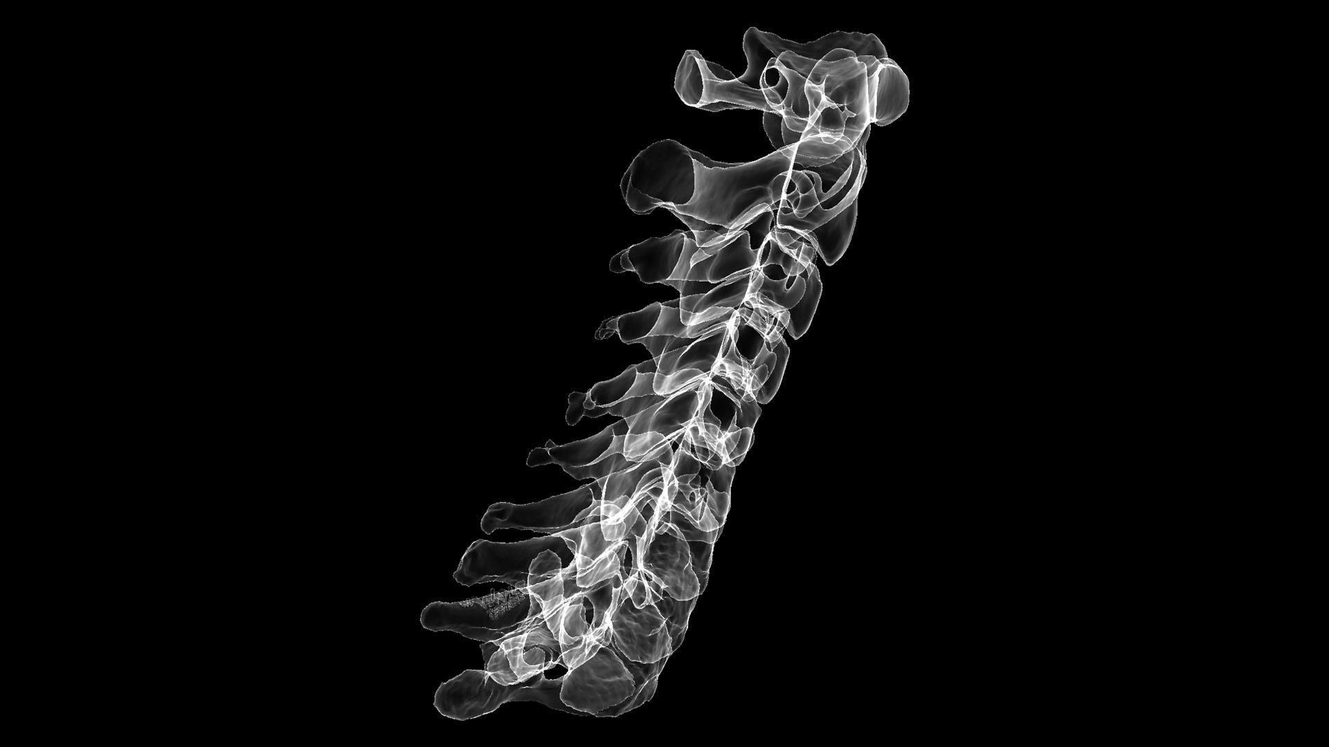
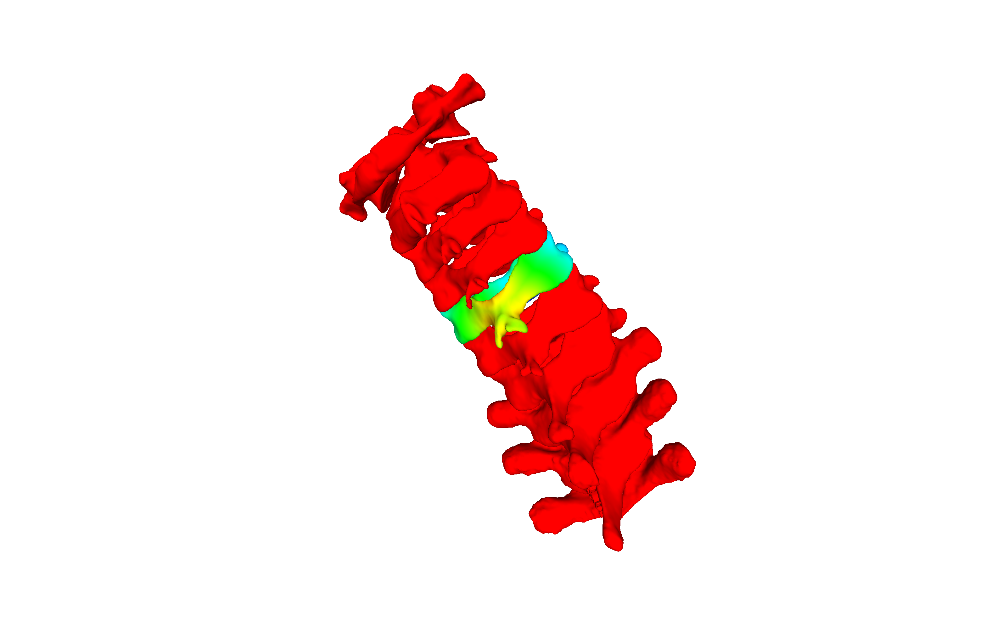
On arrival at the ER, I was separated from my wife and daughter, as only one parent was allowed into the exam room in accordance with COVID policy. The ER doctor ordered a CT of the c-spine and after the radiology team read it we were relieved to learn she had sprained a neck ligament with no signs of anything more serious. My daughter is fine, and other than the general sequelae of having her “bell rung”, she has recovered and is back at school, the incident likely already forgotten. I, on the other hand, still needed to see things for myself. While I can spot the various bones in the 2D DICOM images, to best understand what was going on I wanted to see it in 3D. All we received though, was a cut and pasted finding in the general report.
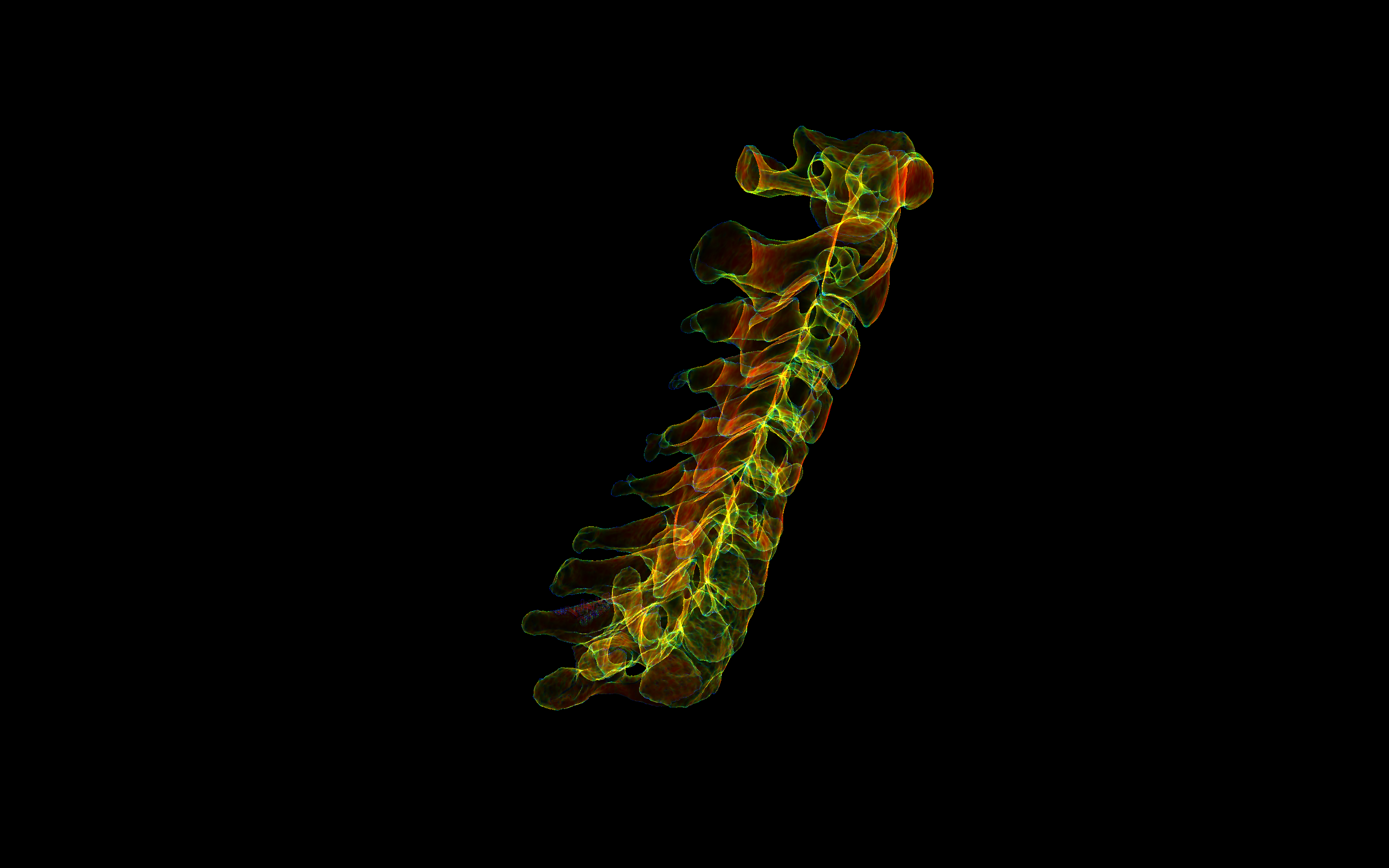
Within radiology departments, 3D views (volume renderings) of scans are routinely available on sophisticated post processing software. For the patient, though, and especially after the reporting is done, getting copies of these 3D files and, more importantly, being able to view them has historically been far from easy. Nowadays, most radiology postprocessing software exports these views as 3D model files suitable for 3D printing. The very same files, however, can easily be viewed on commodity devices such as smartphones and AR/VR headgear. The challenge is to get the file, that is inside the radiology department IT environment, onto one’s viewing device and into a viewer application. This is what we’re facilitating at MedRealilty. So how did it work for my daughter and I?
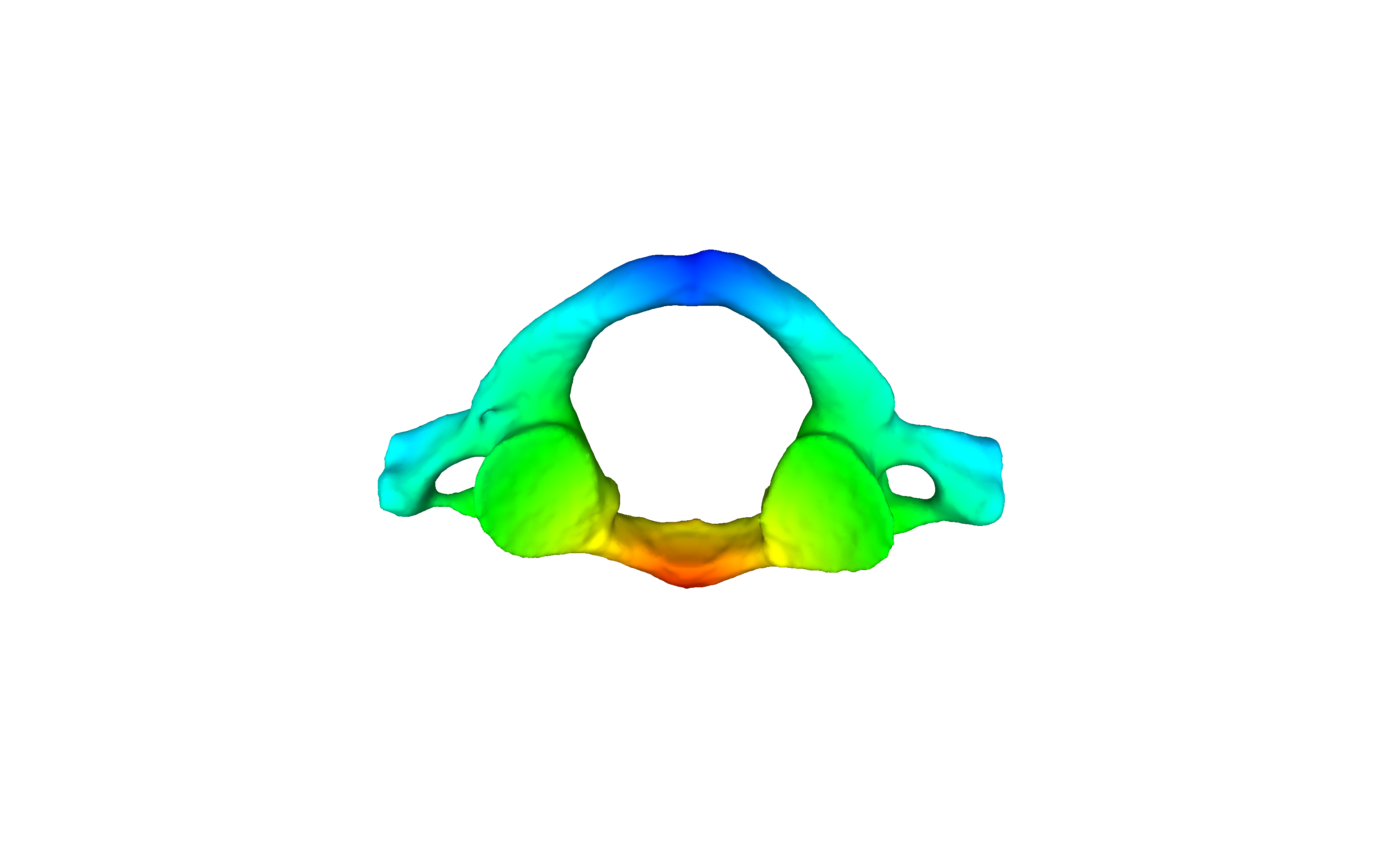
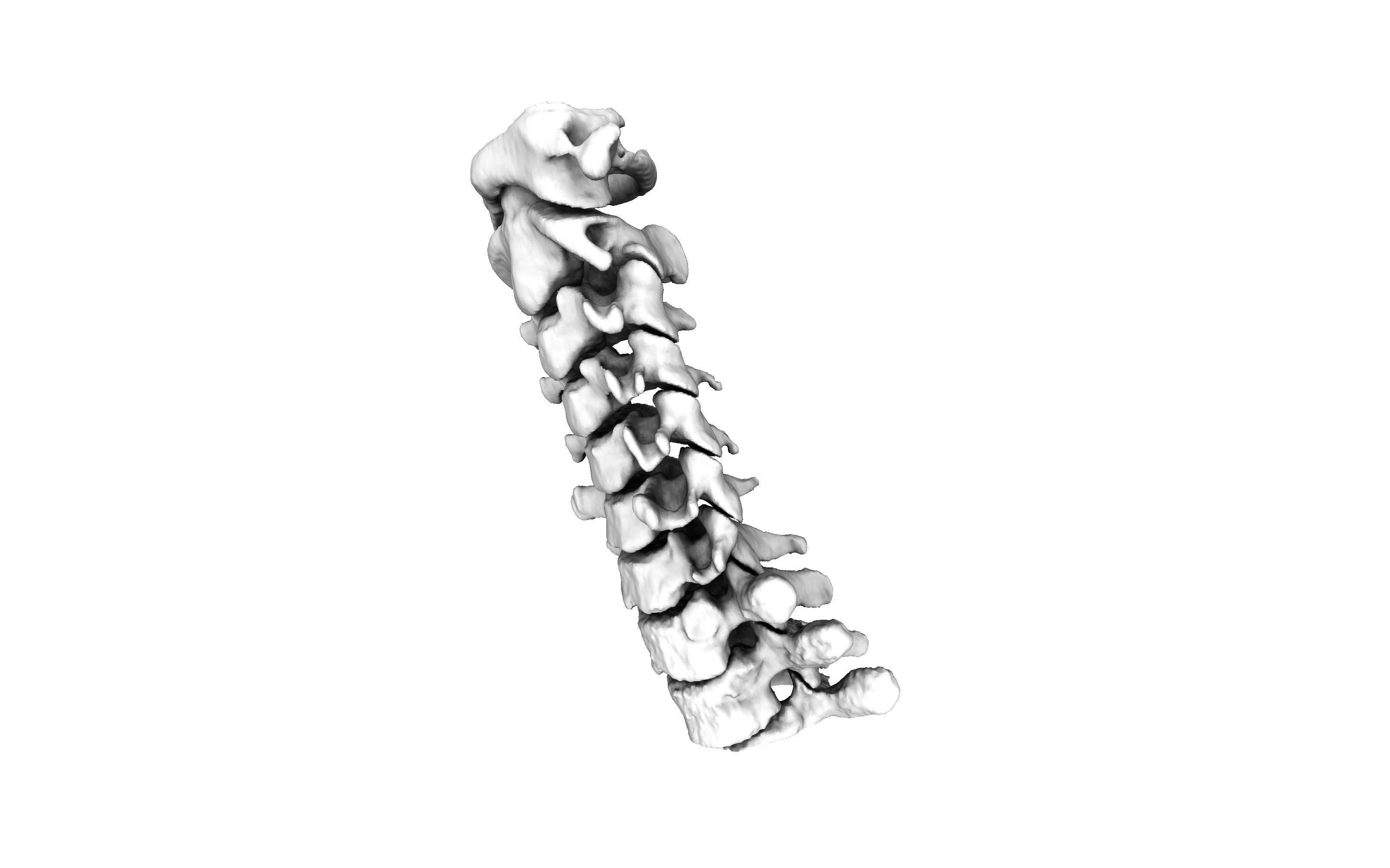
Once I received the DVD from the hospital’s medical records department (requiring filling in some forms and waiting a few days) I still had to get the DICOM transformed into surface models for viewing. For customers who have DICOM studies but need models, MedReality works with a variety of radiology postprocessing companies to have this work done. In my case, Axial3D processed and extracted the surfaces of each cervical vertebra. Once I had the model file in hand, I used the MedReality distribution engine to send it to a variety of devices, including my iPhone, a HoloLens 2 and Oculus Quest. Now I can share these images and explain what happened to her visually to her Mom and to family members across the country.
This is what we’re doing at MedReality: providing access to your volumetric imaging study in a format that the general public can readily understand and on widely available viewing devices. This is how we are changing medicine: by helping radiology become a vibrant part of the general patient experience.
Interested in trying out MedReality? Click here to get started
Share This Story, Choose Your Platform!