Think about your favorite medical drama or criminal-catching series on TV or the big screen; when talking about the human body, do they just use words to describe anatomy to pathology?
No. They don’t.
Hollywood gets the idea: if a picture is worth a thousand words, an interactive 3D model is worth many more. The words they use in the scenes don’t describe the anatomy; most often the dialog is about the insights they are gaining and the ‘a-ha moment’ that helps the case along the path to being solved.
Reality is not as neat, clean and linear as a Hollywood script. In fact, reality is frequently more frightening than even the wildest of imagined scenarios designed for our entertainment. Healthcare is highly personal, complicated, and often confusing to the average person without a medical degree- but it doesn’t have to be.
Across the spectrum of diseases and injuries, determining ‘what is wrong with me’ and ‘how do we fix this’ incorporates non-invasive imaging to provide our caregivers with insight. With over 100 million 3D scans per year across CT, MRI, nuclear medicine, and ultrasound, the world generates more personalized ‘3D content files’ every year than the number of videos uploaded to YouTube since its inception 16 years ago.
So, the digital content exists – but have you ever seen your medical scans presented the way Hollywood creates visual entertainment?
If you are like me, you have not; when I ask for my non-invasive medical data, I get a DVD-ROM with a file called a DICOM (Digital Imaging and Communications in Medicine). When I start up the free viewer usually included with the data from my scan, what I see is the slices through my body, which I have neither the training nor expertise to understand.
Compare the above series of pictures, which are slices through the trunk of the human body. It is difficult for the average person to integrate the slices by visually stacking them in their mind. While we can see the vertebra of the spine, we don’t get a sense of the 3-dimensional nature of our anatomy and pathology.
So, DICOM contains digital imaging, but there is a breakdown in communicating the precise problem, and answering the all-important question: ‘how are we going to thrive?’.
The objective of MedReality is to change this paradigm. Imagine if one of your loved ones was suffering from scoliosis, a deformation of the spine. While the individual slices are critical to your care team for findings, impressions, diagnosis and treatment planning, wouldn’t you prefer to see something more attuned to your level of expertise?
MedReality is a complete ecosystem purposefully designed to bring this 3D data to anyone who needs or wants it in a more immersive, and often less expensive, manner than providing a patient with a DVD-ROM.
We leverage the powerful devices in our pockets, enabling upload of imagery like the spine at left to a mobile phone. We further serve this same content to consumer off-the-shelf augmented reality and virtual reality headsets, 3D displays, and future viewing solutions.
The story of MedReality is intricate – you may be asking where does this content come from, how can I trust it, does it help my care team, does it help me?
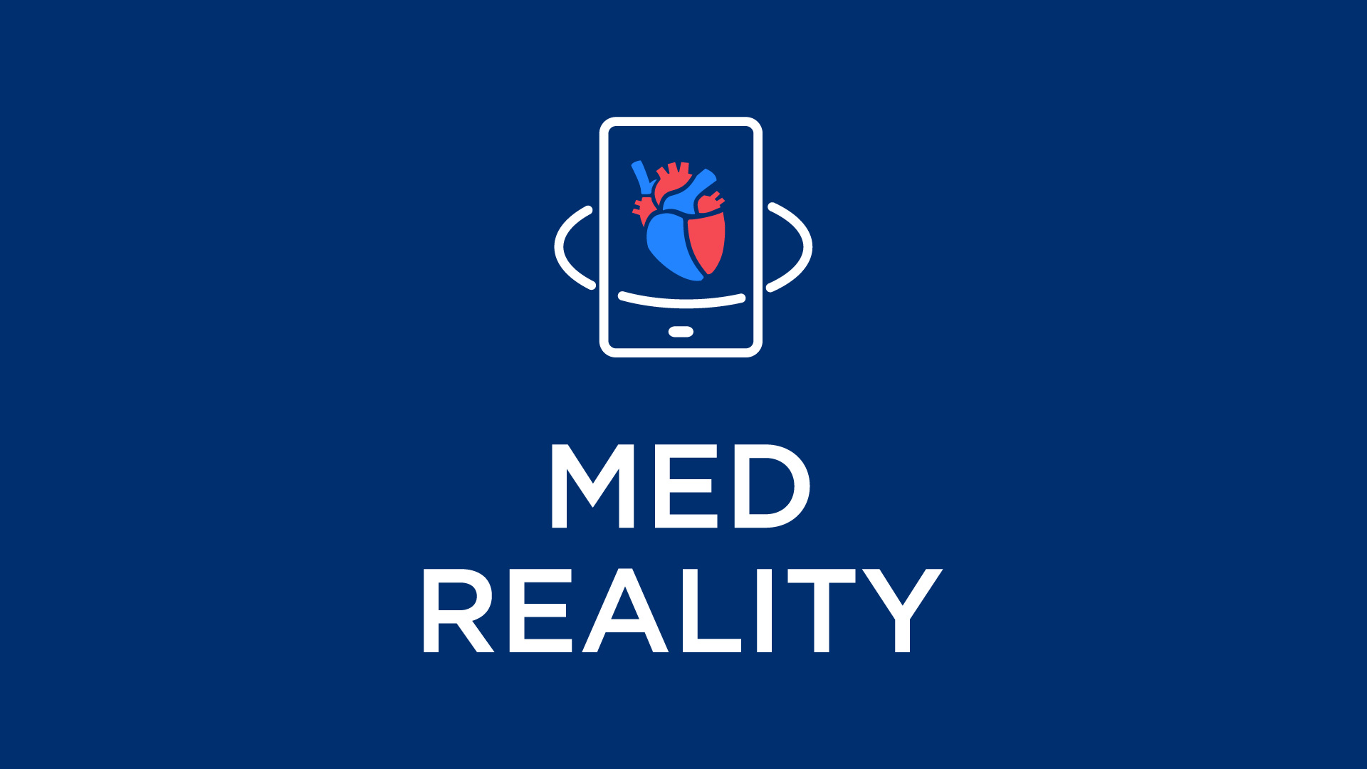
Over the next few weeks we will be introducing MedReality, and answering those questions in addition to exploring some of the topics in the graphic at the top of the page.
Please stay tuned for more posts as we reveal the inner workings of MedReality, how hospitals can use this technology to enhance the patient experience, where to get the content from within the systems your hospital already owns, and many other practical topics.

 (Digital Imaging and Communications in Medicine). When I start up the free viewer usually included with the data from my scan, what I see is the slices through my body, which I have neither the training nor expertise to understand.
 The objective of MedReality is to change this paradigm. Imagine if one of your loved ones was suffering from scoliosis, a deformation of the spine. While the individual slices are critical to your care team for findings, impressions, diagnosis and treatment planning, wouldn’t you prefer to see something more attuned to your level of expertise?